If the CTR is. If you have symptoms such as feeling short of breath a chest x-ray can help doctors find out if its caused by a heart.

Chest Xray Of A Patient Shot In The Chest It S In The Lung And Missed The Heart And Aorta Radiologist Radio Diagnostic Imaging Radiologist Radiology
The heart size should be assessed on every chest X-ray.

Chest x ray heart. For connection of arteries to the aorta angiography would be needed. Chest X-rays can help your doctor determine if there is anything wrong with your heart. What happens during a chest X-ray.
Your doctor uses a chest X-ray to. Assess the hearts borders. Patients who experience symptoms of chest pain a chest injury or shortness of breath.
Chest x ray gives an image of your chest cavity which includes the heart muscle large arteries lungs diaphragm as well as the bones of the rib cage and the spine. Ad Equipped with latest X-Ray Facility and wide range of Medical Check Up Services. EKG Chest X ray can give an impression of thickening of heart muscle or enlargement of heart.
Heart failure occurs when the left ventricular end diastolic pressure rises. On the left a patient with CHF. A CTR of 50 has a sensitivity of 50 for CHF and a specificity of 75-80.
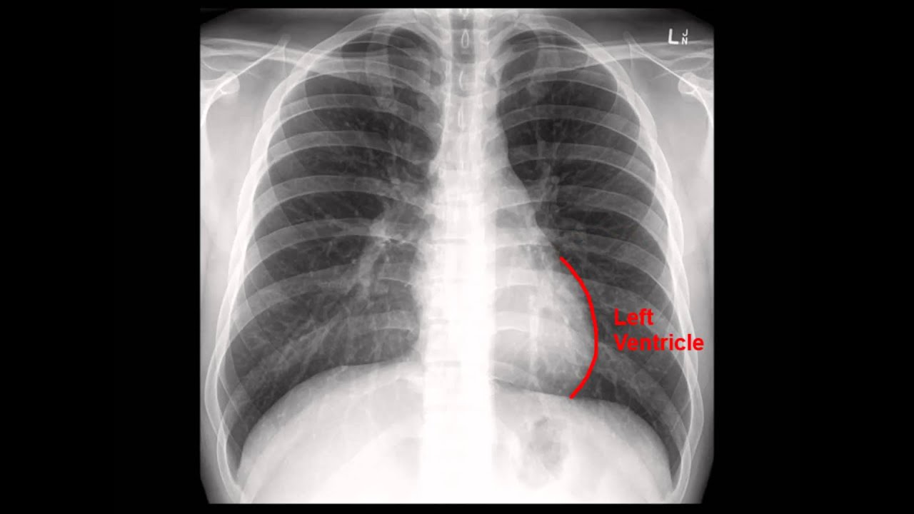
It uses a small amount of radiation to produce an image of your heart lungs and blood vessels. But Echo would be ideal for it. Normal heart appearances as seen on chest X-ray.
One of those is a chest X-ray. In the first phase top right the LVEDP rises above 12 mmHg and on an upright CXR there is equalization of the size of the vessels going to the upper lobes and lower lobes. Ad Find Urgent Care Chest X Ray.
Chest scans can also reveal fluid in or around your lungs or air surrounding a lung. Why is the test done. A chest x-ray produces an image of your heart lungs airways and ribs.
The X-ray technician may ask you to stand still and hold your breath while the images are taken - this helps to provide clearer pictures. It allows doctors to look at your heart lungs and chest wall. There are 3 basic phases of heart failure.
Cardiomegaly can develop for a wide variety of reasons including valvular heart disease cardiomyopathy pulmonary hypertension and pericardial effusion. Although it uses small amounts of ionizing radiations to create an image of the heart muscle it is not the first option when it comes to diagnosing heart. A chest X-ray is a radiology test that creates detailed pictures of your heart lungs andor bones in your chest or spine.
An increase in left ventricular volume of at least 66 is necessary before it is noticeable on a chest x-ray. Another name for a chest X-ray is chest radiograph. During a chest X-ray youll be asked to stand in front of an X-ray machine and press your body against an X-ray plate.
It can help confirm a valve disorder and is useful for diagnosing heart failure or an enlargement of the heart called cardiomyopathy. A chest X-ray can help your doctor see if your heart is an unusual shape or size. However a PA view is required to confidently diagnose cardiac enlargement.
Ad Equipped with latest X-Ray Facility and wide range of Medical Check Up Services. Why might I have a chest x-ray. Learn how to measure the cardiothoracic ratio.
The heart size is considered too large when the CTR is 50 on a PA chest x-ray. Cardiomegaly is said to be present if the heart occupies more than 50 of the thoracic width on a PA chest X-ray. Heart size may be exaggerated by pericardiac fat pads.
Look at your chest bones heart.

Special Investigations In Cardiology Radiology And Electrocardiography Ecg Nurse Radiology Medical

A Future Cardiologist I Have Sent This X Ray Has No Idea What S Wrong Help Him Please Imgur Radiology X Ray Radiology Imaging

Http Www Chestmedicine Org 2015 04 Basics Of Chest X Ray Part 4 Heart And Html X Ray Radiology Medical Knowledge

Chest Xray Shows A Replaced Heart Valve Radiologist Radiology Sob Radiologist X Ray Radiology

X Ray Labels Where The Parts Of The Heart Are Located Click On The Pic On The Left Silhouette Sign Radiology X Ray

Medical School Skin And Bones Xray Art Human

The Radiology Assistant Chest X Ray Basic Interpretation Radiology Radiology Imaging X Ray

Pin On Unusual X Rays

Pin On Comparisons

Chest Radiology Radiology Radiology Student Radiology Technician
Chest X Ray Cxr Analysis In A Nutshell Youtube X Ray Radiology Chest

Pneumonia Bronchitis Bronchitis Pneumonia X Ray

Chest X Ray Heart Failure

Pin Em Medical

By Dr Deepu Basics Of Chest X Ray Part 4 The Heart And Pericardium Radiology Radiology Imaging Cardiac Anatomy

A New Image Everyday With A Description Tricuspid Valve Lung Lobes Mitral Valve
Radiology Chest Xray Normal Radiology Radiology Student Medical Knowledge

Boot Shaped Heart Couer En Sabot Tetralogy Of Fallot Radiology Signs Teaching

Heart And Lungs Chest X Ray Composite Stock Image C011 5758 Xray Art X Ray Heart And Lungs

